Help
Uploads by WikiSysop
Jump to navigation
Jump to search
This special page shows all uploaded files.
File list
Items per page:
20
50
100
250
500
Username:
Include old versions of files
Go
First page
Previous page
Next page
Last page
Date
Name
Thumbnail
Size
Description
Versions
13:23, 27 March 2024
CHECKLIST-LossOfSSEPs.pdf
(
file
)
559 KB
1
20:33, 11 September 2023
Mesenteric Cysts.jpg
(
file
)
31 KB
1
20:31, 11 September 2023
Abdominal Mass Remote Anesthesia.jpg
(
file
)
30 KB
1
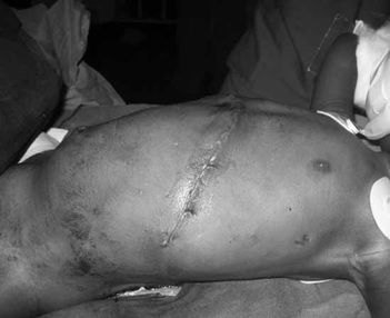
20:29, 11 September 2023
Wilms Tumor Incision.jpg
(
file
)
26 KB
1
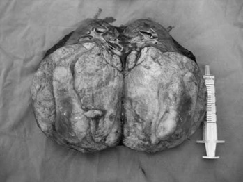
20:27, 11 September 2023
Wilms Tumor Pathology.jpg
(
file
)
29 KB
1
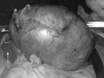
20:26, 11 September 2023
Wilms Tumor Intraoperative.jpg
(
file
)
28 KB
1
20:21, 11 September 2023
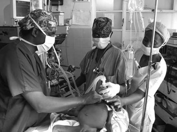
Anesthetic for Wilms.jpg
(
file
)
31 KB
1
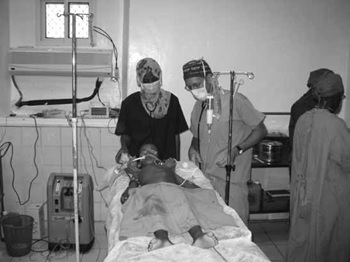
20:05, 11 September 2023
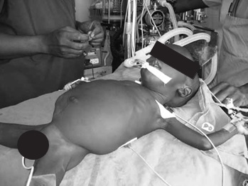
Induction of Child with Large Belly.jpg
(
file
)
38 KB
1
20:02, 11 September 2023
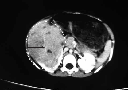
Large Renal Mass2.png
(
file
)
78 KB
1
19:55, 11 September 2023
Large Renal Mass.jpg
(
file
)
18 KB
1
17:32, 7 September 2023
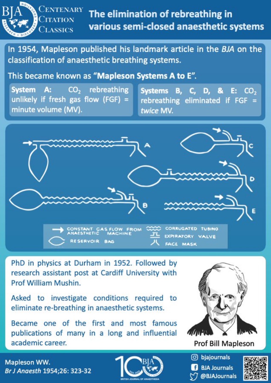
Mapleson Circuits.jpeg
(
file
)
298 KB
1
22:41, 2 July 2023
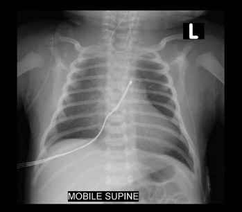
Figure 3 CXR NG tube in pouch.jpg
(
file
)
21 KB
1
22:40, 2 July 2023
Figure 2 Incidence and types of EA TEA.jpg
(
file
)
42 KB
1
22:40, 2 July 2023
Figure 1 Airway an monitoring equipment.jpg
(
file
)
29 KB
1
16:19, 12 February 2023
CICV Algorithm.jpg
(
file
)
156 KB
1
16:19, 12 February 2023
Difficult Tracheal Intubation Algorithm.jpg
(
file
)
193 KB
1
16:19, 12 February 2023
Difficult mask ventilation algorithm.jpg
(
file
)
135 KB
4
23:15, 8 February 2023
LumbarPuntureforSA.jpg
(
file
)
16 KB
1
23:15, 8 February 2023
SpinalNeedlesforSA.jpg
(
file
)
6 KB
1
23:15, 8 February 2023
LateralPositionToPerformSAinNewborn.jpg
(
file
)
23 KB
1
23:15, 8 February 2023
SpinalFormula.jpg
(
file
)
9 KB
1
18:27, 25 February 2022
Hopkins Pedi Anes Pocket Card.pdf
(
file
)
764 KB
1
16:03, 25 February 2022
Hopkins Peds Pocket Card.jpg
(
file
)
1.02 MB
1
15:00, 24 January 2022
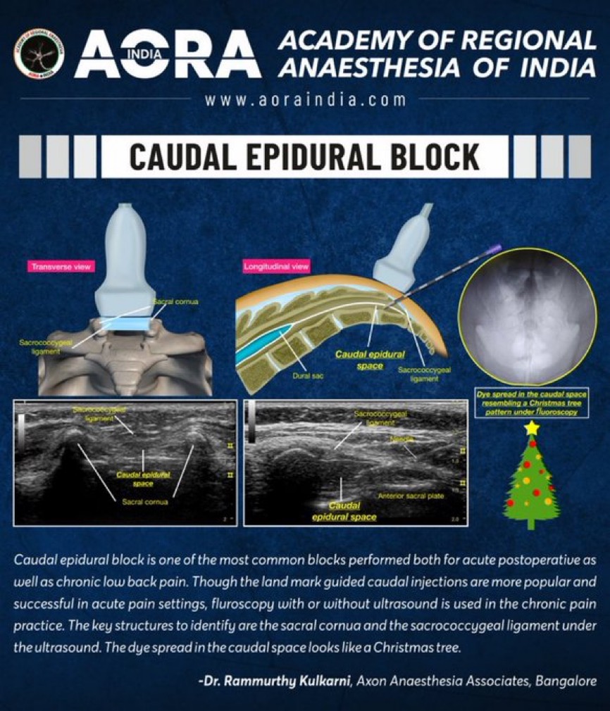
Caudal-Kulkarni.jpeg
(
file
)
257 KB
1
16:31, 2 November 2021
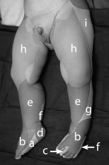
CutaneousNerveSupplyLegFoot.jpg
(
file
)
20 KB
1
16:29, 2 November 2021
AnteriorApproachSciaticNerve.jpg
(
file
)
33 KB
1
16:26, 2 November 2021
PsoasCompartmentBlock.jpg
(
file
)
25 KB
1
16:24, 2 November 2021
AnatomyLumbarSacralPlexus.jpg
(
file
)
30 KB
1
16:22, 2 November 2021
CutaneousNerveSupply.jpg
(
file
)
50 KB
1
16:20, 2 November 2021
AxillaryApproach.jpg
(
file
)
26 KB
1
16:13, 2 November 2021
BrachialPlexus.jpg
(
file
)
27 KB
1
18:49, 30 October 2021
Ultrasound landmarks for TAP block.jpg
(
file
)
19 KB
1
18:47, 30 October 2021
Ultrasound probe position for the transversus abdominis plane.jpg
(
file
)
32 KB
1
18:45, 30 October 2021
Ultrasound landmarks for rectus sheath block.jpg
(
file
)
18 KB
1
18:44, 30 October 2021
Ultrasound probe position for rectus sheath block.jpg
(
file
)
20 KB
1
18:42, 30 October 2021
Injection point for rectus sheath block.jpg
(
file
)
19 KB
1
18:40, 30 October 2021
Ultrasound landmarks for iliinguinal-iliohypogastric.jpg
(
file
)
19 KB
1
18:37, 30 October 2021
Ultrasound probe position for iliinguinal-iliohypogastric.jpg
(
file
)
23 KB
1
18:35, 30 October 2021
Injection point for the ILNB.jpg
(
file
)
30 KB
1
18:33, 30 October 2021
Anatomy of the ilioinguinal-iliohypogastric nerve block.jpg
(
file
)
25 KB
1
16:34, 30 October 2021
Caudal Positive test ECG.jpg
(
file
)
55 KB
1
16:30, 30 October 2021
US use in caudal block.jpg
(
file
)
20 KB
1
16:28, 30 October 2021
US of sacro-coccygeal space.jpg
(
file
)
20 KB
1
16:18, 30 October 2021
Needle misplacement.jpg
(
file
)
63 KB
1
16:16, 30 October 2021
Orientation of the needle during puncture.jpg
(
file
)
18 KB
1
16:13, 30 October 2021
Puncture - orientation of the needle and reorientation after.jpg
(
file
)
36 KB
1
16:10, 30 October 2021
Bony landmarks.jpg
(
file
)
29 KB
1
16:07, 30 October 2021
Preparation of patient - lateral position with the surgical site down1.jpg
(
file
)
23 KB
1
16:03, 30 October 2021
Preparation of patient - lateral position with the surgical site down.jpg
(
file
)
32 KB
1
16:00, 30 October 2021
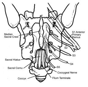
Posterior aspect of sacrum.jpg
(
file
)
34 KB
1
First page
Previous page
Next page
Last page
Navigation menu
Personal tools
English
Create account
Log in
Namespaces
Special page
English
Views
More
Search
Navigation
PedsAnesthesia.Net
PedsAnesthesia Forum
PedsAnesWiki
ABA Keyword List
TYK_Question_List
Help
MediaWiki FAQ
Tools
User contributions
Logs
View user groups
Special pages
Printable version


